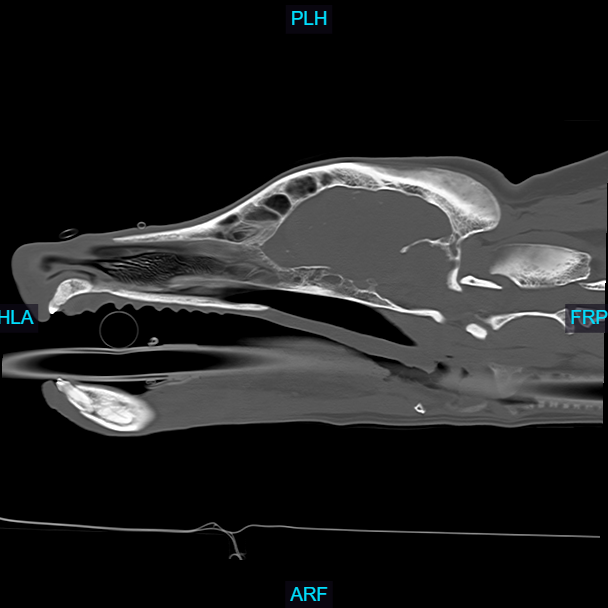
Computed Tomography
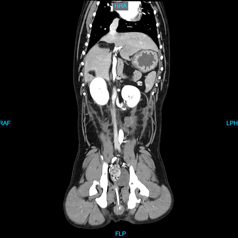
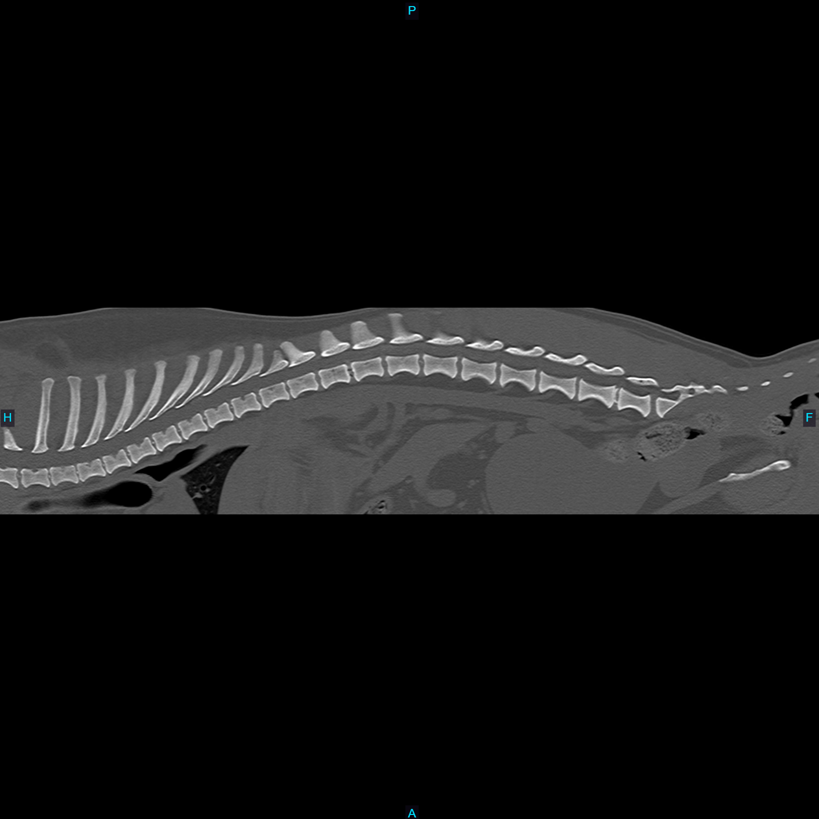
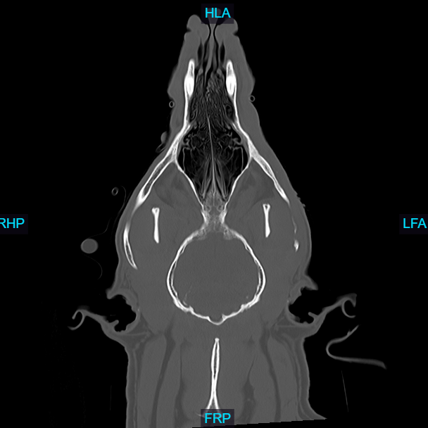
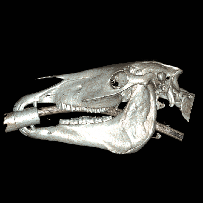
Computed Tomography (CT), also known as a CAT scan, is an advanced imaging technique that combines X-ray technology with computer processing to generate detailed cross-sectional images of an animal's internal structures.
How it works
The animal is typically sedated or anesthetized to remain still during the procedure, ensuring high-quality images. Special positioning aids may be used to maintain the correct posture. The animal is placed on a motorized table that moves through a circular opening of the CT scanner. The scanner consists of an X-ray tube that rotates around the animal, emitting X-rays from different angles. Veterinarians and veterinary radiologists analyze the CT images to identify abnormalities such as tumors, fractures, organ diseases, and other conditions. The high-resolution images allow for a detailed examination that is often not possible with traditional X-rays.
Applications in Veterinary Medicine
- Evaluating complex fractures, joint abnormalities, and spinal disorders.
- Assessing organs like the liver, kidneys, lungs, and intestines for diseases, obstructions,
and other abnormalities.
- Veterinary CT scans are invaluable for providing comprehensive diagnostic information, guiding treatment plans, and improving outcomes for a wide range of animal health issues.

